Scientists can detect defects in graphene thanks to a unique combination of two methods
Graphene has very unique properties and could improve many components and devices. A detailed understanding of the physicochemical properties of this 2D material - including the role of structural defects - is essential for its successful use in practice. Scientists at the J. Heyrovský Institute of Physical Chemistry of the Czech Academy of Sciences have found that by combining two different measurement methods, they can determine the role graphene defects play in transitions between electronic states and electrochemical reactions.
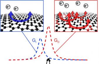
The study was published in The Journal of Physical Chemistry Letters. For the research, the experts used single crystals of graphene tens of micrometres in size. The researchers used so-called microdroplet electrochemistry to transfer electric charge to the basal plane of the graphene sample and measured the corresponding spectral shift using Raman spectroscopy. They noticed that the expected change in the spectral shift depended on the inserted charge and split into two peaks (see figure).

The measured values of the spectral shift at the beginning of the experiment were as expected: the basal plane of the graphene sample seemed to contain no defects (blue curve). However, during the course of the experiment the values started to deviate (red curve).
The experts concluded that the reason for the two peaks in the spectrum is the existence of two different processes with different rates of electric charge transfer. This result is not observable by the Raman spectroscopy alone, which highlights the advantages of combining different experimental methods in-situ.
For practical applications of graphene in electronic devices, batteries or sensors, it is important to know how fast can graphene transfer electrical charge and how this speed relates to defects in the material. Thanks to a unique combination of two different methods, it is now possible to measure these properties.

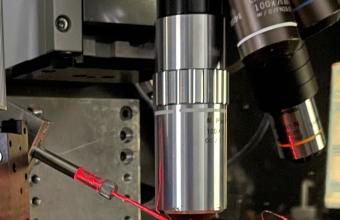
In this way, microdroplet measurements were performed simultaneously with Raman spectroscopy. A red laser-illuminated capillary is used for microdroplet formation, and an objective lens above it is used for visualization of microdroplets and samples and for the Raman spectral measurements.
More information:
Ing. Matěj Velický, Ph.D.
J. Heyrovský Institute of Physical Chemistry of the Czech Academy of Sciences
matej.velicky jh-inst.cas.cz
+420 266 053 755


